胶原纤维、IV型胶原及CD34诊断外伤性迟发性脾破裂
[韩青1, 2  , 阎春霞
, 阎春霞1 , 张毛影1, 3 , 朱杰1 , 顾珊智1, * ]
, 阎春霞]
|
|


第一作者简介:韩青,女,陕西咸阳人,硕士研究生,研究方向为肿瘤的表观遗传学。E-mail: 15529276217@163.com
外伤性迟发性脾破裂的鉴定难点在于准确判断脾破裂与腹部受到外力事件之间是否存在直接延续的因果关系。经典的组织病理检查的结果虽然具有十分重要的作用,但存在假阴性的可能,因此需要寻找其他辅助诊断指标。本文通过一例明确的外伤性迟发性脾破裂病例,在损伤及正常脾组织中,利用HE染色、Masson染色、免疫组化染色,针对胶原纤维、IV型胶原、CD34三个指标进行组织形态学对比,并量化免疫组化结果。HE染色发现出血灶周围已经形成了肉芽组织;Masson染色发现出血区周围的胶原纤维密度明显高于远端正常组织,且形成包膜状纤维结构;免疫组化结果发现,出血区域的IV型胶原及CD34相比正常对照组织均呈强阳性,平均光密度具有统计学差异。在判断脾损伤后经过时间方面,HE染色判断为7~14d,Masson染色判断其大于6d,免疫组化判断为7~14d,三者结果基本一致。说明HE染色、Masson染色以及IV型胶原和CD34免疫组化染色均可获得较为准确的结果。与HE染色相比,胶原纤维、IV型胶原、CD34更为直观并易于识别判断,这为今后此类案件的鉴定提供了新的辅助指标。
Whether there is a direct causal relationship between spleen rupture and accidental abdominal injury is difficult to judge for the identification of traumatic delayed splenic rupture. The approach of classic histopathologic examination, albeit important, lies in possibility of false negative. Thus, the auxiliary diagnostic indicators are helpful and worth seeking. This article deals with a case that has been unambiguously identified as the delayed traumatic splenic rupture. With the damaged and normal (for control) spleen tissues, the manipulations were conducted through the respective HE (hematoxylin-eosin), Masson and immunohistochemical staining to have the three indicators of collagen fiber, collagen IV and CD34 unveiled, therefore making the histological conformation compared together with the quantification of immunohistochemical results. HE staining showed that exuberant granulation had formed around the hemorrhagic foci. Masson staining revealed that the density of collagen fibers was significantly higher around the bleeding area than that of the normal tissue at the distal end, displaying a capsule-like fibrous structure. Immunohistochemical results demonstrated that both the collagen Ⅳ and CD34 were strongly positive throughout the bleeding area when compared with the normal control, exhibiting statistically difference between their average optical densities. On the elapsing time from the injury of spleen, both the HE staining and immunohistochemical results gave 7~14 days, with the Masson’s being not fewer than 6 days, manifesting the roughly same estimation among the three of them for delivery of a comparatively accurate determination. Compared with HE staining, more intuitive and easy-to-recognize results are those indications from collagen fiber, collagen IV and/or CD34, hence the three substances are eligible for the new auxiliary index/indicator into identification of related cases in the future.
外伤性延迟性或迟发性脾破裂(delayed rupture of spleen, DRS)大多数发生在伤后2 周内, 少数发生在伤后4 周内。因其受伤至破裂之间有一定的潜伏期, 故容易引发损伤时间的争议。目前, 法医临床鉴定在判断脾破裂与受伤之间因果关系时, 根据组织损伤后的修复过程具有时序性这一规律, 通常利用HE染色观察脾破裂后不同时间的组织修复的特异性形态改变来判断损伤存在的时间, 其诊断准确性不高。因此, 需要寻找其他辅助诊断指标和更科学客观的鉴定方法。
某男, 14岁, 某年12月21日被同学用膝盖顶伤腹部, 伤后感腹痛, 未就诊。12月21日至28日期间自感腹部隐痛。12月29日至31日腹痛加重, 31日出现恶心呕吐。次年1月1日凌晨患者突感腹部剧痛并晕倒, 送医后诊断为脾破裂, 于当日行脾切除术。现需明确脾破裂性质, 及其与12月21日腹部所受外力之间的因果关系。
B超示脾挫裂伤, 腹腔积液。CT示脾破裂并包膜下血肿, 腹腔积液、积血。
腹腔内有不凝血1 000 mL, 脾肿大, 中下极有一8 cm× 4 cm大小血肿, 中央有一长7 cm、深2 cm的不规则裂口, 周围组织碎裂。切除过程中, 脾内部有黑色血凝块及新鲜出血500 mL, 脾窝后腹膜有血肿。
1.4.1 HE染色
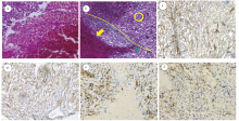
切除的脾组织可见灶片状出血, 出血灶周围可见大量增生的毛细血管和新生的纤维结缔组织, 伴炎细胞浸润, 已经形成了肉芽组织。出血灶周围及远端脾组织呈淤血改变, 但形态结构正常, 无明显病变(图1A)。
 | 图1 脾组织病理学显微镜检照片(A:HE染色〔× 40〕; B:Masson染色〔× 40〕, 胶原纤维呈蓝色〔以曲线为分界, 箭头示脾损伤区, 圆圈示远端正常组织〕; C:损伤区IV型胶原染色〔× 40〕, IV型胶原呈棕色〔阳性〕; D:正常组织IV型胶原染色〔× 40〕, IV型胶原呈棕色〔阳性〕; E:损伤区CD34染色〔× 100〕, CD34阳性细胞呈棕色; F:正常组织CD34染色〔× 100〕, CD34阳性细胞呈棕色)Fig.1 Microscopic observation into histopathologic examination of spleen (A: HE staining (× 40) showing hemorrhage and necrosis of spleen tissue, a large number of proliferated capillaries and new fibrous connective tissue around the bleeding site; B: Masson staining (× 40) revealing collagen fibers shown as blue, with the yellow line as the dividing mark to separate the arrow and circle respectively indicating the splenic damaged area and the distal normal tissue; C/D: collagen IV (× 40) stained of brown as positive in the damaged area/normal tissue; E/F: CD34 cells (× 100) stained of brown as positive in the damaged area/normal tissue) |
1.4.2 Masson染色
脾组织内散在分布呈蓝色的条状纤维样结构, 即为胶原纤维成分。破裂出血区周围的胶原纤维密度明显高于远端正常组织, 且在出血区周围形成包膜状纤维结构(图1B)。
1.4.3 免疫组化染色
1.4.3.1 IV型胶原
脾组织内有大量的条索样、管样不同程度的棕色阳性染色结构。损伤组织中呈强阳性, 且在出血带周围炎细胞渗出带内分布更密集(图1C、1D)。使用IPP软件量化阳性染色强度, 损伤区内平均光密度值(AOD, IOD sum/area sum)为0.42, 正常组织内AOD为0.16, 损伤区域IV型胶原纤维表达明显高于正常脾组织(P< 0.05)。
1.4.3.2 CD34
脾组织内点、线状散在分布呈棕色的CD34阳性细胞。损伤脾组织出血区周围的阳性细胞数量明显多于正常脾组织(图1E、1F)。使用IPP软件量化阳性染色强度, 损伤区内CD34的AOD为0.12, 正常组织内AOD为0.02, 脾损伤区域CD34表达强度明显高于正常脾组织(P< 0.05)。
延迟性脾破裂在外伤发生后, 脾损伤区会发生修复反应, 其过程符合组织修复的一般性表现, 通常会经过三个阶段, 即炎症反应、纤维修复和瘢痕形成。
炎症反应阶段一般为伤后1~2 d, 此时损伤破裂出血引发急性炎症反应, 脾损伤出血区域周围出现中性粒细胞、单核细胞浸润, 并逐渐累积达高峰, 在损伤区周围形成炎细胞聚集带, 并向出血区内迁移, 引起损伤区组织水肿、变性坏死。
纤维修复阶段为伤后3~14 d, 通常在修复早期(伤后3~4 d), 在炎细胞分泌的细胞因子作用下, 损伤区边缘成纤维细胞增生, 新生毛细血管逐渐增多, 开始形成肉芽组织; 随着时间延长, 在伤后5~6 d, 成纤维细胞开始产生胶原纤维; 在伤后7~14 d, 肉芽组织内的炎性细胞、成纤维细胞和毛细血管成分逐渐减少, 胶原纤维数量和体积逐渐增加并达高峰, 至此完成修复阶段。
最终, 在伤后14 d进入瘢痕组织形成阶段, 表现为损伤区域的炎细胞基本消失, 胶原纤维增生停止并开始重排, 毛细血管闭合或改建, 出血区被纤维组织瘢痕取代[1-3]。
本文通过对脾组织HE染色后镜下观察发现, 正常脾组织被出血灶压缩, 出血灶周围有中性粒细胞及纤维素渗出, 部分区域形成含有部分炎细胞的肉芽组织, 符合修复阶段增生中晚期的表现, 判断损伤经过的时间在7~14 d。
为了确定鉴定结果的准确性, 进一步检测修复过程中肉芽组织的分子指标, 包括胶原纤维、IV型胶原及CD34。胶原纤维是肉芽组织的中后期产物, 主要发挥代替原有组织、连接断裂组织的作用。Masson染色可将胶原纤维染成蓝色, 因此利用Masson染色观察胶原纤维分布情况有助于提升判断准确性。
此外, 机体在损伤的诱导下, 来源于骨髓的CD34造血祖细胞被动员进入外周血, 并在趋化因子作用下聚集到创伤组织, 在损伤局部分化为内皮细胞, 形成新血管, 修复损伤[4]。新生的薄壁毛细血管内皮细胞分化成熟后进而分泌IV型胶原等物质构成基底膜的基板。因此, 在组织修复形成的肉芽组织中, CD34与IV型胶原数量不断增多, 分布范围由损伤区域逐渐向外扩大; 随着修复的结束, 两者数量开始减少, 分布范围也不断缩小, 直到瘢痕产生并成熟[5]。
结合本例, 与远端正常组织相比, Masson染色显示脾损伤区域已产生较多胶原纤维, 并且分布较为广泛, 可判断其胶原纤维形成时间大于6 d。同时, IV型胶原染色和CD34细胞染色阳性结果在损伤修复区内分布广泛, 数量较多, 明显高于远离损伤区域的正常脾组织, 可判断其肉芽组织形成已有一段时间但尚未形成瘢痕组织, 脾损伤时间应该处于修复阶段的中晚期, 由此判断损伤时间约为7~14 d。
由此可以看出, 通过胶原纤维、IV型胶原及CD34染色, 可得出与HE染色同样准确的结果, 且更为直观, 易于识别判断。因此得出鉴定结果:伤者脾破裂损伤形成时间范围在7~14 d, 结合调查情况排除其他外力作用因素后, 笔者认为被鉴定人脾破裂与10 d前腹部所受膝顶外力存在因果关系。
综上所述, 本研究结果显示在判断脾组织的损伤时间时, 除了应用传统HE染色外, 还可以进行Masson染色及IV型胶原、CD43免疫组化染色进一步印证HE染色结果, 结合法医临床鉴定中所使用的传统方法, 包括确证外伤史、排除疾病等综合评定, 以期获得更为客观准确的外伤性延迟性脾破裂法医临床鉴定结论。
| [1] |
|
| [2] |
|
| [3] |
|
| [4] |
|
| [5] |
|